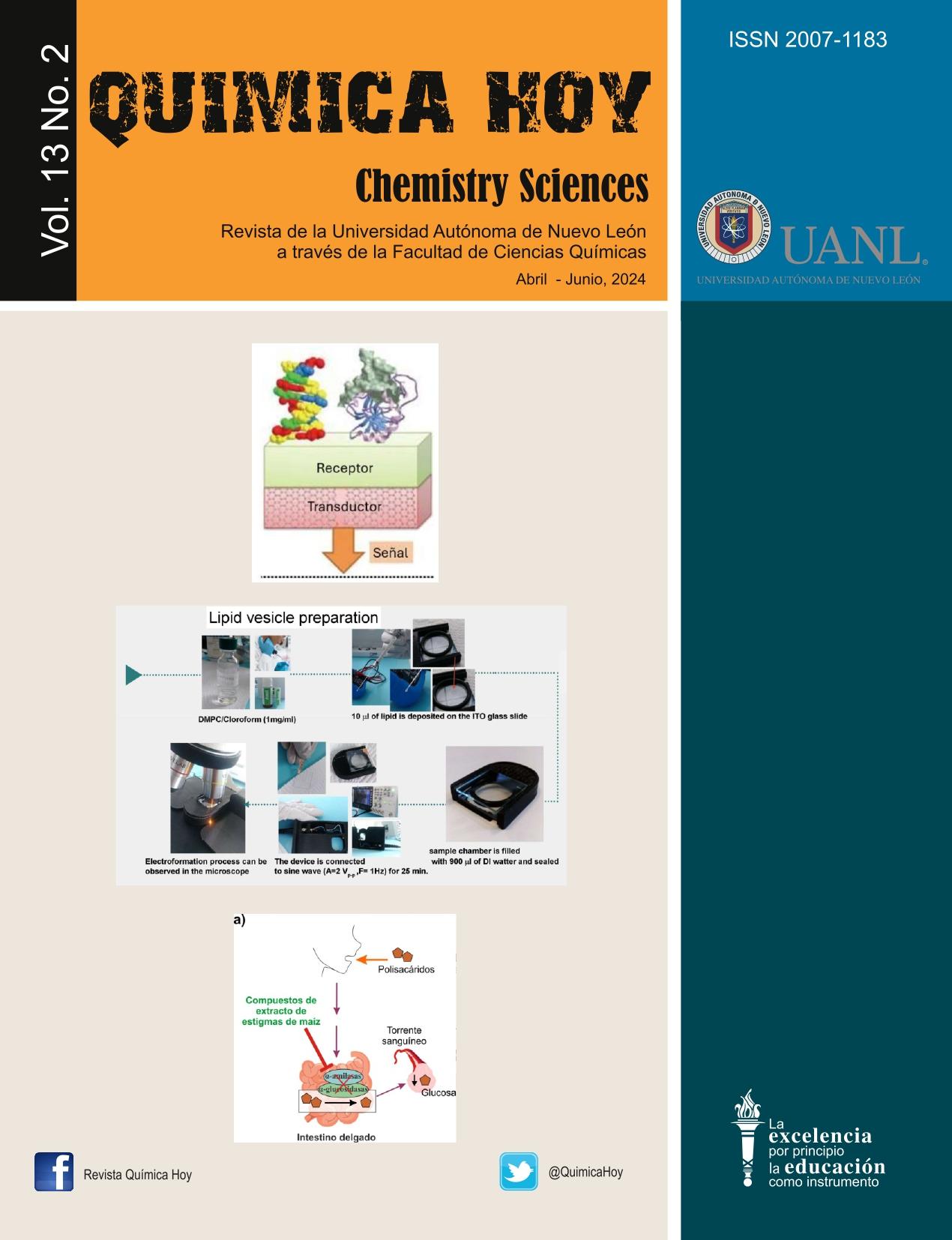
A Versatile DIY 3D Printed Device for the Preparation of Gian Lipid Vesicles as Model Membranes by Electroformation: Design and Fabrication
DOI:
https://doi.org/10.29105/qh13.02-412Palabras clave:
3D Printing, Giant Vesicles, Phospholipids, CAD-CAM, Model MembranesResumen
Una forma sencilla de estudiar las propiedades de la membrana celular y su interacción con otras moléculas es mediante el uso de sistemas modelo con la misma estructura básica. Estos utilizan unos pocos componentes básicos que simulan las condiciones de la membrana plasmática y se han utilizado ampliamente para estudiar las propiedades de las membranas biológicas. Entre estos sistemas modelo, podemos encontrar mono capas de Langmuir, bicapas soportadas y vesículas lipídicas. Las vesículas lipídicas son particularmente interesantes debido a que su estructura esférica imita a las membranas celulares. Entre los diferentes procedimientos para preparar GUV's (vesículas unilamelares gigantes), la electroformación destaca porque las vesículas producidas por este método son en su mayoría más grandes que 100 nm y unilamelares, lo que las hace fácilmente visibles mediante microscopía óptica convencional. Aunque existen dispositivos comerciales disponibles, generalmente se fabrican equipos personalizados de manera artesanal. Este trabajo describe el diseño y la fabricación de un dispositivo sencillo y versátil impreso en 3D para preparar vesículas lipídicas gigantes utilizando el método de electroformación. El dispositivo ha sido diseñado para fijarse en la platina del microscopio.
Descargas
Citas
- [1]. Kurniawan J, Yin NN, Liu GY, Kuhl TL. Interaction forces between temaiy lipid bilayers containing cholesterol. Langmuir 2014;30:4997-5004. https://doi.org/10.1021/la50034lc. DOI: https://doi.org/10.1021/la500341c
- [2]. Dimova R,Aranda S,Bezlyepkina N, Nikolov V, Riske KA, Lipowsky R. A practical guide to giant vesicles. Probing the membrane nanoregime via optical microscopy. J Phys Condens Matter 2006;18. https://doi.org/10.1088/0953- 8984/18/28/S04. DOI: https://doi.org/10.1088/0953-8984/18/28/S04
- [3]. Subczynski WK, Wisniewska A. Physical properties of lipid bilayer membranes: Relevance to membrane biological functions. Acta Biochim Pol 2000;47:613-25. https://doi.org/10.18388/abp.2000_3983. DOI: https://doi.org/10.18388/abp.2000_3983
- [4]. Veatch SL, Keller SL. Separation of Liquid Phases inGiant Vesicles of Ternary Mixtures of Phospholipids and Cholesterol. Biophys I 2003;85. https://doi.org/10.1016/S0006-3495(03)74726-2. DOI: https://doi.org/10.1016/S0006-3495(03)74726-2
- [5]. Van Meer G, Voelker DR, Feigenson GW. Membrane lipids: where they are. Nat Rev Mol Cell Biol 2009;101:1-4. https://doi.org/10.1038/nrm2330.Membrane.
- [6]. Stefaniu C, Brezesinski G, Möhwald H.Langmuir monolayers as models to study processes at membrane surfaces. Adv Colloid Interface Sci 2014;208:197-213. https://doi.org/10.1016/j.cis.2014.02.013.
- [7]. Veatch SL, Cicuta P, Sengupta P, Honerkamp-Smith A, Holowka D, Baird B. Critical fluctuations in plasma membrane vesicles. ACS Chem Biol 2008;3. https://doi.org/10.1021/cb800012x. DOI: https://doi.org/10.1021/cb800012x
- [8]. Karslake J, Stone M, Veatch SL. Probing Sub-Micron Critical Composition Fluctuations using Super-Resolution Techniques in Giant Plasma Membrane Vesicles. Biophys J 2014;106. https://doi.org/10.1016/j.bpj.2013.11.583. DOI: https://doi.org/10.1016/j.bpj.2013.11.583
- [9]. Veatch SL, Keller SL. Seeing spots: Complex pase behavior in simple membranes. Biochim Biophys Acta - Mol Cell Res . 2005;1746 https://doi.org/10.1016/j.bbamcr.2005.06.010 DOI: https://doi.org/10.1016/j.bbamcr.2005.06.010
- [10]. Bañuelos-Frías A, Castañeda-Montiel VM, Alvizo-Paez ER, Vazquez-Martinez EA, Gomez E, Ruiz-Garcia J. Thermodynamic and Mechanical Properties of DMPC/Cholesterol Mixed Monolayers at Physiological Conditions. Front Phys 2021;9:1-10. https://doi.org/10.3389/fphy.2021.636149. DOI: https://doi.org/10.3389/fphy.2021.636149
- [11]. Brezesinski G, Möhwald H.Langmuir monolayers as models to study processes at membrane surfaces. Adv Colloid Interface Sci 2003;208:197—213. https://doi.org/10.1016/j.cis.2014.02.013. DOI: https://doi.org/10.1016/j.cis.2014.02.013
- [12]. Méléard P, Bagatolli LA, Pott T. Giant Unilamellar Vesicle Electroformation. From Lipid Mixtures to Native Membranes Under Physiological Conditions. Methods Enzymol2009;465:161-76. https://doi.org/10.1016/S0076- 6879(09)65009-6. DOI: https://doi.org/10.1016/S0076-6879(09)65009-6
- [13]. Moscho A, Orwar OWE, Chiu DT, Modi BP, Zare RN. Rapid preparation of GUVs. Proc Natl Acad Sci USA 1996;93:11443-7. DOI: https://doi.org/10.1073/pnas.93.21.11443
- [14]. Angelova M, Dimitrov DS. A mechanism of liposome electroformation. Trends Colloid Interface Sci II 2007:59-67.https://doi.org/10.1007/bfb0114171. DOI: https://doi.org/10.1007/BFb0114171
- [15]. Brownell WE, Jacob S, Hakizimana P, Ulfendahl M, Fridberger A, Jargensen IL, et al. Termaiy pase diagram of dipalmitoyl-PC/dilauroyl-PC/cholesterol: Nanoscopic domain formation driven by cholesterol. Biophys J 2013;8:234-67. https://doi.org/10.1007/s00249-016-1155- 9.
- [16]. Estes DJ, Mayer M. Giant liposomes in physiological buffer using electroformation in a flow chamber. Biochim Biophys Acta - Biomembr 2005;1712:152-60. https://doi.org/10.1016/j.bbamem.2005.03.012. DOI: https://doi.org/10.1016/j.bbamem.2005.03.012
- [17]. Kuribayashi K, Tresset G, Coquet P, Fujita H, Takeuchi S. Electroformation of giant liposomes in microfluidic channels. Meas Sci Technol 2006;17:3121-6. https://doi.org/10.1088/0957-0233/17/12/S01 . DOI: https://doi.org/10.1088/0957-0233/17/12/S01
- [18]. Diegel O, Nordin A, Motte D. Additive Manufacturing Technologies. 2019. https://doi.org/10.1007/978-981-13- 8281-9_2. DOI: https://doi.org/10.1007/978-981-13-8281-9_2
- [19]. Ngo TD, Kashani A, Imbalzano G, Nguyen KTQ, Hui D. Additive manufacturing (3D printing): A review of materials, methods, applications and challenges. Compos Part B Eng 2018;143:172-96. https://doi.org/10.1016/j.compositesb.2018.02.012. DOI: https://doi.org/10.1016/j.compositesb.2018.02.012
- [20]. Yazdi AA, Popma A, Wong W, Nguyen T, Pan Y, Xu J. 3D printing: an emerging tool fornovel microfluidics and labon-a-chip applications. Microfluid Nanofiuidics 2016;20:1-18. https://doi.org/10.1007/s10404-016-1715-4. DOI: https://doi.org/10.1007/s10404-016-1715-4
- [21]. blender.org. blender.org -Home of the Blender project - Free and Open3D Creation Software. Blenderorg 2015.
- [22]. Ultimaker. Cura 3D Printing Slicing Software. Ultimaker 2017.
- [23]. Simons K, Vaz WLC. Model systems, lipid rafts, and cell membranes. Annu Rev Biophys Biomol Struct 2004;33:269-95. https://doi.org/10.1146/annurev.biophys.32.110601.14180 3. DOI: https://doi.org/10.1146/annurev.biophys.32.110601.141803
- [24]. Shitamichi Y, Ichikawa M, Kimura Y. Mechanical properties of a giant liposome studied using optical tweezers. Chem Phys Lett 2009;479:274-8. https://doi.org/10.1016/j.cplett.2009.08.018. DOI: https://doi.org/10.1016/j.cplett.2009.08.018
- [25]. Wu H, Yu M, Miao Y, He S, Dai Z, Song W, et al. Cholesterol-tuned liposomal membrane rigidity directs tumor penetration and anti-tumor effect. Acta Pharm Sin B 2019;9:858-70. https://doi.org/10.1016/j.apsb.2019.02.010. DOI: https://doi.org/10.1016/j.apsb.2019.02.010
- [26]. Bertrand B, Munusamy S, Espinosa-Romero IF, Corzo G, Arenas Sosa I, Galván-Hernández A, et al. Biophysical characterization of the insertion of two potent antimicrobial peptides-Pin2 and its variant Pin2[GVG] in biological model membranes. Biochim Biophys Acta - Biomembr 2020;1862. https://doi.org/10.1016/j.bbamem.2019.183105. DOI: https://doi.org/10.1016/j.bbamem.2019.183105
- [27]. Witkowska A, Heinz LP, Grubmüller H, Jahn R. Tight docking of membranes before fusion represents a metastable state with unique properties. Nat Commun 2021;12. https://doi.org/10.1038/s41467-021-23722-8. DOI: https://doi.org/10.1038/s41467-021-23722-8
- [28]. Jergensen IL, Kemmer GC, Pomorski TG. Membrane protein reconstitution into giant unilamellar vesicles: a review on current techniques. Eur BiophysJ 2017;46. https://doi.org/10.1007/s00249-016-1155-9. DOI: https://doi.org/10.1007/s00249-016-1155-9
- [29]. Hernández-Adame PL, Meza U, Rodríguez-Menchaca AA, Sánchez-Armass S,| Ruiz-García J, Gomez E. Determination of the size of lipid rafts studied through single-molecule FRET simulations. Biophys J 2021;120. https://doi.org/10.1016/j.bpj.2021.04.003. DOI: https://doi.org/10.1016/j.bpj.2021.04.003
- [30]. Wesolowska O, Michalak K, Maniewska J, Hendrich AB. Giant unilamellar vesicles -a perfect tool to visualize phase separation and lipid rafts in model systems. Acta Biochim Pol 2009;56:33-9. https://doi.org/20091772 [pii]. DOI: https://doi.org/10.18388/abp.2009_2514
Descargas
Publicado
Cómo citar
Número
Sección
Licencia
Derechos de autor 2024 Alan Bañuelos Frías, Claudia Valero Luna, Athziri Herrera Saucedo, Cristina Flores Cadengo, Pablo Luis Hernandez Adame, Francisco Bañuelos Ruedas, Lazaro Canizalez DavaIos, Leo Alvarado Perea, Alfonso Talavera Lopez

Esta obra está bajo una licencia internacional Creative Commons Atribución 4.0.


